

To celebrate National Science Week, 12 August – 20 August, EducationDaily is publishing a series of STEM-focused articles featuring inspiring Australians and innovative ideas.
It’s mystical and mesmerising, and presents fascinating scientific art from some of Australia’s most creative and visionary researchers.
The unique Art of Science competition and exhibition is back in 2023 and features pieces by people who spend their working days trying to solve the world’s most complex medical challenges.
This innovative annual art show enables viewers to explore the latest biomedical research through a range of captivating still and moving digital images that connect science and art in colourful, creative ways.
Researchers from the Walter and Eliza Hall Institute of Medical Research (WEHI) shows the world of scientific research as one of beauty and wonder – and help curious minds discover the powerful story behind each image.
Intricate biological details, such as a growing nerve cell, blood vessels in the lung, or even milk-producing cells in the breast, reveal a hidden inner-world normally only seen in laboratories, through a microscope.
Art of Science has a historical background dating back to 1997, when the competition was created by former WEHI director Professor Suzanne Cory.
This year, the launch of the online gallery of the 16 still and moving images chosen as the finalists in WEHI’s 2023 Art of Science competition – with winners judged by The Age and Sydney Morning Herald’s national science reporter Liam Mannix – coincides with National Science Week (12-20 August) and shines a spotlight on WEHI’s research, from understanding the drivers of cancer and malaria, to unravelling the mysteries of cell death.
The 2023 Art of Science will be WEHI director Professor Doug Hilton’s last under his leadership, with Professor Hilton appointed the next Chief Executive of the Commonwealth Scientific and Industrial Research Organisation (CSIRO), commencing on 29 September.
“In over three decades at WEHI and 14 years as director, I have been privileged to watch Art of Science grow and to witness the relentless passion, dedication and innovation of our researchers,” Professor Hilton said.
“WEHI’s research aims to ensure more people can live healthier for longer. Along the way, we want to share the beauty and wonder of our work and reveal to the world the intricate complexities of pioneering medical research. Through their creativity, our scientists inspire the future generations that will continue to drive the research and discovery we need to solve our most critical health challenges,” he said.
Building upon WEHI’s successful collaboration with the Melbourne Conservatorium of Music at the University of Melbourne last year, and the overwhelmingly positive response received, we are delighted to announce our renewed partnership in 2023.
To learn more, EducationDaily asked three of the WEHI researchers turned digital artists about their professional passion and how they find art in science.
‘Into the Matrix’ – Raymond Qin and Niall Geoghegan (music by Margaret Wozniak)
Raymond Qin’s inspiration to enter this year’s Art of Science competition comes from a passion for science and communication.
“The Art of Science competition offers me a unique platform to promote complex scientific concepts in beautiful imagery,” he told EducationDaily. “It’s an honour be featured amongst these amazing scientists/artists.”
With the exhibition aiming to demonstrate that “rigorous scientific research can produce artistic patterns, forms, and processes”, Mr Qin hopes hope this exhibition will spark interest in students and inspire young scientists to develop a career in science.

“Having our work featured means that the work we do can have a positive impact to the science community and society, enabling a broader audience to see and enjoy,” he says. “Our work also highlights the interdisciplinary collaboration between me and Dr. Niall Geoghegan, showcasing the strong connection between different departments at WEHI.”
Immune cells need to move through a range of body tissues to hunt out and kill infected or cancerous cells. But many organs and tissues contain fibres made of collagen, a protein found throughout the body, particularly in skin, bones, teeth, ligaments, tendons and other connective tissues.
Using a lattice light-sheet microscope, researchers Mr Qin and and Mr Geoghegan have been able to show, in 3D, how an immune cell (magenta) contorts into different shapes to find the best route through the dense matrix of collagen fibres (grey).
A better understanding of how immune cells navigate is crucial in helping researchers understand the immune response and find new treatments for a range of diseases.
‘Disco’ – Sabrina Lewis (music by Sadie Mustoe)
When Sabrina Lewis was in her high school biology class, watching an animation created by a biomedical animations specialist from WEHI, she immediately found learning the process of DNA replication so much easier to visualise and understand – and motivated her own professional pathway.
Today, the work Ms Lewis does in the imaging lab complements WEHI’s broader focus on visualising biological processes at a microscopic level to aid medical breakthroughs.
By using powerful microscopes to take high-resolution images of cancer cells, Ms Lewis says “our hope is that understanding how the cancer cells behave and what genes they are expressing might reveal novel therapeutics”.

“Science is inherently creative,” she told EducationDaily. “Whether it’s designing new experiments to answer biological questions or capturing beautiful images under a microscope. I hope this exhibition displays some of the creativity and beauty we see every day in the lab, and sparks conversations at home or even inspires a new generation of scientists.”
Cancer tissues are not uniform, and visualising where specific molecules and biomarkers are located within a cancer can provide insights as to where and when drugs might be used more effectively to target cancers and treat disease.
PhD student, Ms Lewis, has used fluorescent molecular labels and confocal microscopy to produce a “map” of molecules within cancer cells in a small region of a tumour. This moving image takes us through a forest of shimmering lines, each representing the location of a specific molecule as visualised from side-on.
Techniques like this that map spatial variability within tumours – so-called “spatial omics” – are expected to be important in developing the next generation of diagnostic and therapeutic strategies for cancer.
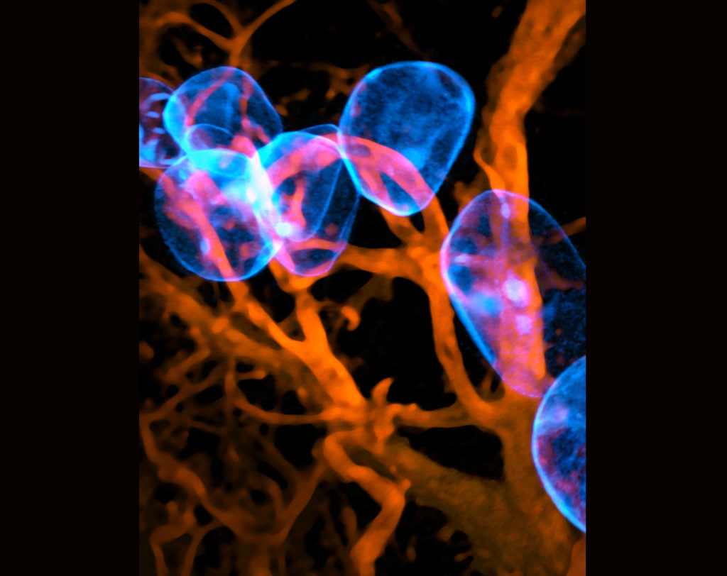
‘Oh it Glows, Adipose’ – Caleb Dawson
“I have the privilege of using fluorescence microscopy in my research on breast immunology, which produces images like this,” Caleb Dawson told EducationDaily. “Every day, I see the incredible shapes and patterns of the cells in our organs, and I always have an urge to share this with others. As a Christian, these microscopic worlds give me a glimpse of the power and artistry of God, which is my main inspiration in science.”
“The Art of Science, says Mr Dawson, is “a great celebration of life, beauty and truth, as well as the ground-breaking research that the artworks come from”.
“It’s always a joy to be a part of a brilliant exhibition like this, so I’m grateful for being selected.”

“Life at every scale, Mr Dawson told EducationDaily, “is a wonderfully elaborate work of art, so it’s inevitable that science will keep uncovering art that amazes us and could belong in a museum”.
“I hope this competition shows many people a beautiful aspect of life that they would never have considered or imagined before,” he says.
While trying to view milk-producing cells of the breast, researcher Caleb instead stumbled on these adipose cells, lit up like glowing blue orbs by fluorescent proteins.
The breast adipose cells – otherwise known as fat cells – are 30-40 micrometres in size, about the diameter of a human hair. They are surrounded by red blood vessels that weave throughout the breast tissue.
Cells are normally impossible to differentiate from one another, as they are packed closely together and have no intrinsic colour. But by lighting them up with fluorescent proteins, researchers are better able to understand the roles different cells play in organ function and disease, knowledge that can then be applied to creating and improving treatments.
And the winners are…
First place in the 2023 Art of Science competition still image category went to Heart of the Storm by Farzaneh Shojaee.
Judge and journalist Liam Mannix called it “a visually striking image that draws one’s attention instantly, and then as you look closer only becomes more and more detailed”.
In Heart of the Storm, all looks calm and serene in this section of intestinal wall. But closer examination reveals that a ‘storm’ has passed through, leaving damaged and dead cells in the finger-like villi that extract essential nutrients from digested food.
Haemophagocytic lymphohistiocytosis (HLH) is a severe and often fatal syndrome in which a viral infection or other illness triggers the immune system to become overactive and attack healthy cells.
PhD student Mr Shojaee is investigating pathways responsible for inflammation and cell death in HLH, with the aim of identifying potential new treatments.
“Intestinal wall showing damage from haemophagocytic lymphohistiocytosis (HLH), a severe and often fatal syndrome in which infection or other illness activates the immune system to attack healthy cells.”
The winner in the moving images category is Hanadi Hoblos’ Four Seasons (music by Nicholas Dullow).
This work turns living cells into dancers that float and sway on the breeze, dancing together or alone. Beautiful, elegant, and captivating,” Mr Mannix said.
Every living cell has a dynamic and continuously changing “cytoskeleton” made up of microtubules and actin filaments that give the cell its shape, help organise its parts, and provide a basis for movement and cell division.
Honours student, Mr Hoblos, has used lattice light sheet microscopy to show the remarkable abilities of living cells to reorganise and change their form.
DCLK1 is highly expressed in a range of cancers and may be involved in the ability of cancer cells to migrate and invade normal tissues, making it a potential target for the development of cancer treatments.
Other award winners in 2023 are:
- Oh it Glows, Adipose by Caleb Dawson (Second Place – Still Image)
- On Fire by Claire Marceaux and Aysha Al-Ani (Third Place – Still Image)
- Disco by Sabrina Lewis (Second Place – Moving Image)
- Imagined Protein by Lachlan Whitehead, Marjan Hadian-Jazi and Richard Birkinshaw (Third Place – Moving Image)